
 Medical imaging is one of the many informational diagnostic tools for healthcare providers to review to inform your comprehensive medical care. Whether you have previous imaging reports or are hoping to have imaging performed, we are able to help you navigate this process. Below, we will discuss the basics of different types of imaging and how Bissell Clinic can help you learn about your anatomy and respond to your symptoms.
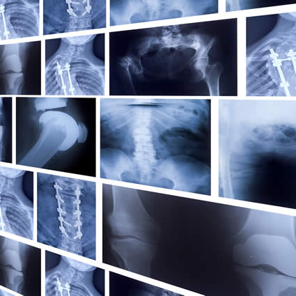
X-Ray
When most people think of having x-rays taken, they are referring to radiography, which is the least expensive, quickest and most common form of medical imaging we will discuss here. Other types of x-rays include CT (computed tomography) scans and fluoroscopy. These three types of imaging are considered x-ray technology because images are formed by x-rays passing through the body and sending information to a receiver reflecting absorption and dispersion across different tissues.
Radiography
This is the most common application of x-ray technology. Radiographs capture a single image to be processed and reviewed after the imaging is completed. Radiographs are best for detecting joint pathology such as narrowed joint space as well as dislocations and fractures. Mammograms also fall under the radiography category.
CT
CT exams essentially function as a compilation of many single x-ray images to create a 360 degree view of internal structures. Patients often get CT exams done in emergency situations because these exams take very little time and provide detailed results. CT exams can detect blood clots and soft tissue injuries as well as fractures or other structural disruptions that may or may not be visible on x-rays.
Fluoroscopy
Flouroscopy is used for real-time visualization of tissue. Applications include for monitoring an interventional procedure such as a spinal injection or the passage of contrast dye throughout body tissues.
MRI
Magnetic resonance imaging (MRI) is another non-invasive form of medical imaging which uses a large magnet to pass radiowaves through tissue. This allows for extremely-detailed visualization of soft tissue pathology such as cartilage loss, spinal injuries, nerve disruption, inflammation, and ligament, tendon, or bone disruption. MRIs do not expose the patient to radition of any kind and allow healthcare providers to sift through subtleties that go undetected with x-ray imaging. Depending on the size of the magnet, MRIs may take between 10-30 minutes to complete.
DEXA
Dual-energy x-ray absorptiometry (DEXA) is used to measure bone density and determine location and severity of bone loss. This is the most common way to diagnose osteoporosis and is a quick, non-invasive study.
Blood Testing
Blood testing is a great tool for diagnosing and management of interal diseases or processes including inflammation, infection, rheumatoid arthritis, gout, thyroid function, and electrolyte imbalances, among many others. Blood work can also be helpful in identifying nutrient deficiencies and for guidance in micronutrient supplementation.